Understanding Synovial Cysts in the Spine: A Detailed Overview
Intro
Synovial cysts are fluid-filled sacs that can develop around the joints, and they may also occur in the spinal region. These cysts often arise in relation to facet joint osteoarthritis or other pathologies affecting the spine. As interest grows in understanding the impact of these cysts on spinal health, it becomes vital to explore their clinical relevance, treatment options, and implications for overall patient care.
Overview of Research Topic
Brief Background and Context
Synovial cysts form when synovial fluid leaks from a joint space and accumulates, forming a cyst. In the spine, these cysts typically appear in the lumbar region. The cysts may cause symptoms by exerting pressure on nearby spinal structures, leading to pain, weakness, or other neurological deficits. The growing recognition of their potential for serious complications necessitates a deeper examination into their formation and management.
Importance in Current Scientific Landscape
The understanding of synovial cysts in the spine influences both diagnosis and treatment strategies. As more research emerges, it highlights the need for precise identification and tailored therapeutic approaches, making this an important topic in current medical discourse. Information gained from recent studies can aid medical practitioners in improving patient outcomes. The exploration of this topic can also deepen comprehension of spinal health in general, offering valuable insights for educators and researchers alike.
Methodology
Research Design and Approach
This overview is based on an extensive review of current literature surrounding synovial cysts in the spine. Key studies, case reports, and systematic reviews provide a solid foundation for understanding the nature of these cysts, their origins, and solutions to manage them effectively. A broad interpretation of both clinical and laboratory findings ensures a multifaceted perspective on this condition.
Data Collection Techniques
Data was collected through various methods, including:
- Literature reviews from medical journals and databases.
- Interviews and surveys conducted with healthcare professionals who specialize in spinal disorders.
- Case studies, which provide insights into real-life scenarios and treatment outcomes.
The integration of various sources of information helps to paint a comprehensive picture of the condition, revealing insights that are crucial to advancing the understanding of synovial cysts in the spine.
Prelims to Synovial Cysts
Synovial cysts are a common yet often misunderstood condition related to spinal health. Understanding these cysts is critical for medical professionals, researchers, and patients alike. They may contribute to significant discomfort and can impact the overall quality of life.
Cysts generally may form near the synovial joints. These specific cysts develop due to the abnormalities in the surrounding synovial tissue, leading to a fluid-filled sac. Their presence is typically linked to underlying spinal conditions, such as arthritis or trauma. By recognizing their characteristics and implications, practitioners can provide better care for patients experiencing symptoms.
Definition and Characteristics
A synovial cyst is defined as a fluid-filled sac that arises from the synovial tissues. These tissues line the joints, secreting synovial fluid that lubricates and nourishes the joint space. Synovial cysts vary in size, and their structure can change over time. They are typically unilocular, meaning they contain a single chamber filled with synovial fluid.
Characteristics include:
- Location: Commonly linked to the lumbar spine but can occur in other regions.
- Size: Ranging from a few millimeters to several centimeters in diameter.
- Symptoms: Patients may experience pain, stiffness, or neurological symptoms if the cyst compresses nearby structures.
These cysts can be benign, but they may also complicate existing spinal issues.
Historical Context
The understanding of synovial cysts has progressed over decades. Earlier reports focused primarily on their occurrence without emphasizing the underlying pathophysiological mechanisms. Advances in imaging technology, such as MRI, have allowed for better visualization of these structures, changing the approach towards diagnosis and treatment.
In the 20th century, healthcare professionals began recognizing the association between spinal arthritis and cyst formation. This led to a deeper understanding of how synovial cysts could arise from degeneration in joints. Recent studies have aimed to establish clearer connections between these cysts and other spinal disorders, emphasizing the need for continued research in this area.
"Knowledge of synovial cysts is essential, as these entities can significantly impact both diagnosis and treatment plans in spinal care."
Understanding the historical context enhances the appreciation for current research. It also paves the way for future developments in treatment options and diagnostic criteria.
Anatomy of the Spine
Understanding the anatomy of the spine is crucial when discussing synovial cysts. The spine serves as the central support structure in the human body. It protects the spinal cord and provides stability. An understanding of spinal anatomy helps in recognizing where synovial cysts typically form and their potential impact on surrounding structures.
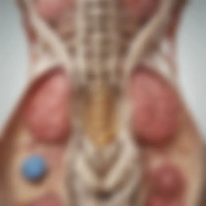
The spine is divided into several regions: cervical, thoracic, lumbar, sacral, and coccygeal. Each region has distinct characteristics, important for diagnosing and treating related conditions. Synovial cysts are predominantly found in the lumbar region, where the mobility and load-bearing nature create an environment conducive to their formation.
Spinal Structure Overview
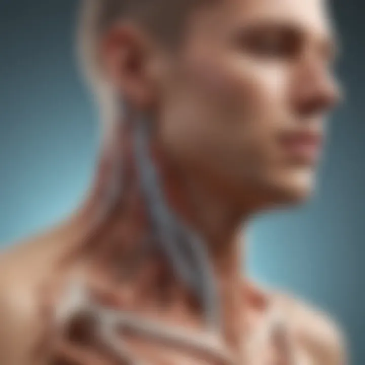
The spinal column consists of vertebrae, intervertebral discs, ligaments, and surrounding muscles. Vertebrae stack upon each other, creating a column that houses the spinal cord. This structure protects vital nervous tissue but is also susceptible to mechanical stress.
- Vertebrae: 33 individual bones organized into five regions. The lumbar vertebrae are larger, to bear more weight.
- Intervertebral Discs: Act as shock absorbers, providing cushioning between vertebrae. They have a nucleus pulposus at the center and an annulus fibrosus surrounding it.
- Ligaments: Provide stability, connecting different vertebrae together. The anterior longitudinal ligament prevents hyperextension, whereas the posterior longitudinal ligament protects against hyperflexion.
- Muscles: Surrounding muscles facilitate movement and maintain posture.
This anatomy is essential in the discussion of synovial cysts, as these cysts originate near synovial joints in the lumbar region, impacting surrounding structures.
Role of Synovial Joints
Synovial joints play a significant part in spine mobility. These joints allow for a greater range of movement. They are lined with synovial membrane, which produces synovial fluid. This fluid nourishes and lubricates the joint surfaces. In the context of synovial cysts, these joints are the primary sites of cyst formation.
The role of synovial joints includes:
- Facilitating Movement: These joints allow bending and rotation of the spine.
- Providing Stability: While allowing mobility, they also contribute to overall spinal stability.
- Cushioning: The synovial fluid acts as a cushion, reducing wear on the joints during movement.
Dysfunction or irritation in these joints can lead to the formation of cysts. Understanding the mechanics of synovial joints is important. It equips medical professionals to address the implications of these cysts more effectively
"The insights gained from studying the anatomy of the spine provide foundational knowledge essential for understanding the complexities associated with synovial cysts."
In summary, a comprehensive understanding of spinal anatomy lays the groundwork for addressing the clinical implications of synovial cysts. This knowledge is vital for researchers and medical practitioners involved in spinal health.
Pathophysiology of Synovial Cysts
The pathophysiology of synovial cysts in the spine is a crucial aspect of understanding this condition. Synovial cysts generally form due to some mechanical stress or injury. They originate from synovial membrains which are present in the joints and are responsible for lubricating them. When these membranes become disrupted, due to trauma or degenerative changes, they can begin to protrude and form cysts. The cysts often contain synovial fluid, which adds to the complexity of the condition.
Another important factor is the relationship between synovial cysts and other spinal conditions, such as degenerative disc disease. Lack of stability in the spine could lead to the development of these cysts. One must consider the intricate dynamics of spine biomechanics while studying the origin and implications of synovial cysts.
Formation Mechanism
The formation mechanism of synovial cysts begins with instability or degeneration in the surrounding joints. It can happen when a joint undergoes excessive wear and tear, leading to potential injury of the synovial lining. When the synovial lining is damaged, it may produce an excessive amount of fluid. This fluid can collect in spaces, forming the cyst. Over time, this process may create significant pressure on nearby structures, such as nerve roots and spinal cord.
Key points in this mechanism include:
- Trauma: Sudden injury can initiate the formation.
- Degenerative Conditions: Diseases like osteoarthritis increase the risk.
- Joint Synovium: Excess fluid accumulation leads to cyst development.
Biomechanical Factors
Biomechanical factors play a central role in synovial cyst formation. When the spine experiences abnormal movement patterns or stress, it can contribute to changes in the cartilage and the synovial membrane. Over time, these stressors can lead to microtrauma, setting the stage for cyst development.
The loading of the spine during daily activities can affect how forces are distributed across the joints. If the joint is overloaded, it may lead to structural changes and promote synovial fluid accumulation. Moreover, the alignment of the spine can either alleviate or exacerbate these issues.
Understanding the biomechanical factors can also aid in prevention and treatment strategies. Clinicians may focus on addressing alignment and stability through physical therapy to mitigate risks.
Thus, examining the biomechanical context fosters a comprehensive understanding of how synovial cysts emerge and impact spinal health.
In summary, comprehending the pathophysiology of synovial cysts encompasses the study of both their formation mechanism and biomechanical factors. This exploration sheds light on their clinical significance and could guide future management approaches.
Symptoms and Clinical Presentation
The discussion of symptoms and clinical presentation is central to understanding synovial cysts in the spine. These cysts can lead to various clinical signs that, if properly identified, may assist in timely diagnosis and management. Awareness of these symptoms allows healthcare professionals to differentiate between related spinal conditions, guiding them toward appropriate treatment strategies. Knowing the common symptoms and their neurological implications is crucial for effective patient management and improving quality of life for affected individuals.
Common Symptoms
Synovial cysts in the spine can result in a wide range of symptoms, though not all individuals will experience the same issues. Here are some common symptoms associated with these cysts:
- Localized Pain: Patients often report pain in the area of the cyst, which can vary in intensity. This pain may be sharp or dull and often radiates into neighboring regions.
- Stiffness: Many individuals may experience stiffness in their back or neck. This discomfort can limit mobility and impact daily activities.
- Weakness: Muscle weakness may occur, particularly if the cyst compresses nearby nerves.
- Sensory Changes: Alterations in sensation, such as numbness or tingling, can manifest in the arms or legs, depending on the location of the cyst.
- Radiculopathy: Patients may experience symptoms of radiculopathy, which include pain that radiates along the course of a nerve.
These symptoms not only indicate the presence of a synovial cyst but also highlight the importance of subsequent diagnostic evaluation to establish a precise diagnosis.


Neurological Implications
The neurological implications of synovial cysts are significant. As these cysts develop, they may exert pressure on adjacent spinal nerves or the spinal cord itself. This pressure can cause serious neurological deficits. Consider the following implications:
- Nerve Compression: Compression of spinal nerves can lead to motor and sensory disturbances. Patients might notice an inability to control certain movements or loss of sensation in specific areas.
- Progressive Symptoms: The presence of a cyst can lead to progressive symptoms over time. If untreated, the resulting neurological involvement can worsen, necessitating more urgent intervention.
- Quality of Life Impact: The neurological symptoms associated with synovial cysts can profoundly affect a patient's quality of life. Chronic pain and reduced mobility can create substantial emotional and psychological challenges.
It is vital for practitioners to recognize the range of neurological implications these cysts can bear on a patient’s overall health. Prompt diagnosis and management can mitigate these risks.
In summary, understanding the symptoms and their neurological implications is essential for appropriate clinical management. Awareness can lead to timely interventions that improve the outcomes for individuals living with synovial cysts in the spine.
Diagnosis of Synovial Cysts
Diagnosis of synovial cysts in the spine is crucial for effective management and treatment planning. Accurate diagnosis helps differentiate synovial cysts from other similar lesions and conditions, ensuring that appropriate therapeutic interventions are applied. Delays or inaccuracies in diagnosis can lead to significant complications, affecting a patient's overall health and quality of life. Understanding how to properly identify these cysts is therefore paramount in spinal care.
Radiological Techniques
Radiological techniques are central to diagnosing synovial cysts. The most commonly used imaging modalities include:
- Magnetic Resonance Imaging (MRI): This technique is highly prized for its ability to provide detailed images of soft tissues. Synovial cysts appear as well-circumscribed lesions with high signal intensity on T2-weighted images. MRI allows for the assessment of the cyst’s size, location, and any impact on surrounding structures, such as nerve roots.
- Computed Tomography (CT) Scans: CT scans offer a comprehensive view of bony structures and can highlight bone erosion or other changes caused by the cyst. They are particularly useful in cases where MRI is contraindicated or unavailable.
- Ultrasound: Although not commonly used for spinal cysts, ultrasound can play a role in guiding injections or biopsy when necessary. Its advantage lies in being a real-time imaging technique that does not involve ionizing radiation.
Utilizing these techniques provides a nuanced understanding of the cyst’s characteristics, aiding healthcare professionals in making informed diagnoses.
Differential Diagnosis
Differentiating synovial cysts from other spinal lesions is essential. Several conditions can mimic the presentation of synovial cysts, including:
- Ganglion Cysts: Often located near joints, they can present similarly but are usually found outside the spinal canal.
- Herniation of Intervertebral Discs: Disc herniation can compress nerve roots and present similar symptoms. Identifying the source of nerve compression is vital.
- Tumors: Benign or malignant tumors can appear as cystic lesions on imaging, making histological evaluation necessary in ambiguous cases.
- Infection: Conditions such as abscesses or other infectious processes can also present as fluid-filled lesions. Clinical history and lab tests play important roles in making this distinction.
By thoroughly evaluating these possibilities, healthcare professionals can ensure a precise diagnosis and avoid mismanagement of the underlying condition. The integration of proper diagnostic techniques and a clear understanding of differential diagnoses is essential in achieving optimal outcomes in spinal health.
Management Options
Effectively managing synovial cysts in the spine is crucial for maintaining spinal health and preventing further complications. The management strategies can broadly be divided into non-surgical and surgical interventions. Understanding these options allows medical practitioners to tailor treatment to individual patient needs, maximizing recovery outcomes and enhancing quality of life.
Non-surgical Approaches
Non-surgical management focuses on conservative strategies aimed at alleviating symptoms and improving functionality without the need for surgical intervention. These methods can be particularly beneficial for patients who may not yet require surgery or are not suitable candidates for such procedures. Key non-surgical options include:
- Physical Therapy: Tailored physical therapy programs can help strengthen the muscles surrounding the spine, improving stability and reducing pressure on cysts. A physical therapist can recommend specific exercises that promote flexibility, strength, and pain relief.
- Medications: Non-steroidal anti-inflammatory drugs (NSAIDs) can reduce inflammation and alleviate pain associated with synovial cysts. Corticosteroids may also be injected directly into the cyst to decrease swelling and discomfort.
- Observation: For asymptomatic cysts, a conservative approach may involve regular monitoring. This method allows healthcare providers to assess whether the cyst changes in size or affects spinal function over time.
- Lifestyle Modifications: Encouraging patients to adopt ergonomic practices can help limit strain on the spine, reducing symptoms. Activities like frequent breaks during prolonged sitting can help minimize discomfort.
Deciding on non-surgical options should be based on the patient's overall health, the severity of symptoms, and the cyst's characteristics.
Surgical Interventions
When conservative treatment options fail to relieve symptoms or when the cyst leads to significant neurological issues, surgical intervention may be warranted. Surgical options aim not only to remove the cyst but also to address associated complications. Common surgical procedures include:
- Cyst Excision: This is the most direct approach, involving the complete removal of the cyst. Success rates for this procedure are typically high, leading to symptom relief in many patients. However, the complexity may vary depending on the cyst's location.
- Laminectomy: This surgery involves removing part of the vertebra to relieve pressure on spinal nerves. It may be performed in conjunction with cyst excision if the cyst’s location has caused nerve compression.
- Microsurgical Techniques: Advancements in microsurgery enable surgeons to perform minimally invasive procedures with less tissue damage and quicker recovery times. These techniques often lead to less postoperative pain and shorter hospital stays.
- Fusion Surgery: In cases where spinal instability is indicated, fusion surgery might be considered. This procedure joins two or more vertebrae to stabilize the spine.
Surgical options require thorough evaluation and should involve patient discussions about risks and benefits. It is crucial to consider individual patient profiles before determining the most suitable approach.
Post-Treatment Outcomes
Post-treatment outcomes are a critical aspect when managing synovial cysts in the spine. Understanding the results of treatment helps practitioners evaluate the effectiveness of interventions and gauge the cyst's implications on individual patient health. The recovery process and long-term prognosis provide invaluable insights into the challenges patients may face after treatment.
Recovery Process
The recovery process following treatment for synovial cysts can vary significantly among patients. It largely depends on the specific treatment used, whether surgical or non-surgical, as well as the patient's overall health and the severity of their condition.
- Surgical Recovery: For those who undergo surgical intervention, recovery often involves a few phases. The initial post-operative phase may be marked by swelling and discomfort. Physical therapy is usually recommended to enhance mobility and strength. This can be crucial for the complete restoration of function. The timeline for return to normal activities can range from several weeks to months, depending on the extent of the surgery.
- Non-Surgical Recovery: In cases where non-surgical methods are utilized, such as injections or other conservative measures, the recovery can often be quicker. Patients may experience gradual improvement, but it is important to adhere to recommended follow-ups to monitor the cyst’s behavior.
- Monitoring: Regardless of the treatment approach, ongoing monitoring is essential to assess the cyst's status. Regular imaging tests can provide insight into whether the cyst is shrinking, stable, or potentially recurring. This will guide further management if necessary.
Long-term Prognosis
The long-term prognosis for individuals treated for synovial cysts in the spine is generally favorable but can vary based on several factors.
- Recurrence Rates: Some studies suggest that there is a possibility of recurrence, particularly in cases where conservative treatments were ineffective. Surgical removal generally has lower rates of recurrence compared to non-surgical options.
- Functional Outcomes: Most patients report an improvement in symptoms and overall spinal function post-treatment. However, some may continue to experience lingering issues such as mild pain or reduced range of motion. This can impact daily routines and quality of life.
- Long-term Monitoring: It is recommended that patients engage in long-term follow-up care. This aids in early detection of potential complications or recurrences. Continued assessment ensures that any issues can be addressed promptly.
"Awareness of the potential challenges and benefits of treatment can empower patients in their recovery journey and lead to better management of their spinal health."
Complications Associated with Synovial Cysts
Understanding the complications associated with synovial cysts in the spine is critical for comprehensively addressing this condition. These complications can impact patient health, treatment approaches, and overall well-being. It is essential for healthcare providers, researchers, and educators to have a thorough knowledge of these associated risks and their implications in clinical practice.
Potential Risks
Synovial cysts may lead to several potential risks that require attention. Among the foremost concerns is the risk of nerve compression. As these cysts grow, they can exert pressure on nearby neural structures. This may result in neurological symptoms such as pain, muscle weakness, or sensory disturbances. Additionally, cysts can cause spinal instability when located in regions vital for structural support.
Some possible complications include:
- Infection: Though rare, there is a chance of cyst infection, leading to severe complications such as an abscess.
- Hemorrhage: In cases where cysts rupture, there may be bleeding, which can complicate the patient's clinical picture.
- Recurrence: Even after surgical removal, synovial cysts may reappear, necessitating further interventions.
Impact on Quality of Life
The presence of synovial cysts can significantly influence an individual's quality of life. Individuals suffering from symptomatic cysts may experience chronic pain and limited mobility. This can lead to difficulties in maintaining daily activities, affecting work productivity and social interactions. The emotional toll cannot be understated either; living with pain can result in anxiety and depression.
Research indicates that:
- Patients report a decreased ability to participate in recreational activities.
- Chronic pain can lead to sleep disturbances, exacerbating fatigue and irritability.
Current Research and Advances
The study of synovial cysts in the spine has seen significant progress in recent years. Understanding current research and advances is key for healthcare professionals and researchers alike. This knowledge can lead to better diagnosis, treatment, and management options for patients suffering from these spinal conditions. Ongoing studies focus on the etiology, pathophysiology, and effective treatment protocols, reflecting a comprehensive approach to spinal health.
Recent Findings
Recent studies have revealed insights that enhance the understanding of synovial cysts. For instance, researchers have identified specific biomarkers that could help in differentiating between malignant and benign cysts. The use of advanced imaging techniques, such as high-resolution MRI, has improved the visualization of these cysts, allowing for more accurate diagnoses.
Some findings indicate a possible link between the mechanical stress on the spine and the formation of these cysts. Increased activity in osteoarthritic joints may lead to cyst formation, suggesting that monitoring joint health could be essential in prevention strategies. More studies also emphasize the importance of a multidisciplinary approach in managing synovial cysts, bringing together orthopedic surgeons, neurologists, and rehabilitation specialists.
Future Directions in Research
As understanding of synovial cysts evolves, future research must address several areas. One critical area is the exploration of minimally invasive treatment options, as several studies suggest that surgery may not always be necessary. Ongoing research into the role of biologics and regenerative medicine in treating these cysts could be transformative.
There is also a pressing need to understand the genetic factors associated with synovial cyst formation. Identifying genetic predispositions may help in developing targeted therapies, potentially reducing the occurrence of these cysts in high-risk populations.
In summary, the current research on synovial cysts in the spine emphasizes the importance of interdisciplinary collaboration. It promises advancements in treatment methodologies that are less invasive and more effective. Continued investigations into the causes, effects, and best treatment practices will contribute significantly to the field of spinal health.
"Advancements in our understanding will lead to more effective and personalized care for individuals with synovial cysts."
By staying informed about these developments, professionals can better address the complexities related to synovial cysts in clinical settings.
End
The conclusion serves a vital role in synthesizing the extensive information presented about synovial cysts in the spine. It encapsulates the critical findings from the article while reinforcing the relevance of understanding these particular cysts in the broader context of spinal health.
Summary of Key Points
In reviewing the key points about synovial cysts, several important considerations arise:
- Definition and Formation: Synovial cysts arise from the development of synovial fluid-filled sacs in the spine, often linked to degenerative changes in the spinal joints.
- Symptoms: Typical symptoms include localized pain and potential neurological issues, depending on the cyst's location and size.
- Diagnosis: Advanced imaging techniques, such as MRI and CT scans, are essential for accurate diagnosis.
- Treatment Options: Management can range from non-surgical methods, like physical therapy, to surgical interventions for symptomatic relief.
- Complications and Quality of Life: Understanding the complications associated with these cysts is crucial, especially how they may impact the patient's quality of life.
Call for Continued Research
The complexity of synovial cysts necessitates ongoing research to improve diagnostic accuracy and treatment efficacy. Continued exploration in the following areas is recommended:
- Mechanisms of Formation: Understanding the underlying mechanisms that lead to the formation of these cysts could open new avenues for prevention and treatment.
- Long-term Outcomes: Researching the long-term outcomes for patients treated for synovial cysts will aid in developing best practices in management.
- Innovative Treatment Modalities: Case studies and clinical trials on novel therapeutic approaches should be conducted to assess efficacy and safety comprehensively.
As medical science progresses, these efforts will ultimately contribute to enhanced care strategies for individuals affected by synovial cysts in the spine.
