Understanding the Causes of Pericardial Effusion


Intro
Pericardial effusion refers to the buildup of fluid within the pericardial cavity, the thin sac surrounding the heart. This condition can lead to various complications if not addressed timely. The dynamics of pericardial effusion merit serious attention due to its potential impact on cardiac function. To navigate through these complexities, one must first grasp the underlying causes that contribute to this medical issue.
This article delves into a range of etiological factors that orchestrate the occurrence of pericardial effusion, spanning infectious agents such as viruses and bacteria, autoimmune diseases leading to systemic inflammation, malignancies which may produce exudate, and idiopathic cases whose origins remain elusive. Understanding these causes provides a crucial foundation for effective diagnosis and treatment.
The exploration of this condition sheds light on the delicate interactions within the cardiovascular system. To further elaborate on this subject, we will discuss common diagnostic techniques as well as various management strategies, resulting in a comprehensive overview of pericardial effusion and its implications.
Overview of Research Topic
Brief Background and Context
Pericardial effusion can arise from a multitude of pathophysiological processes. It has been noted that the accumulation of fluid may be attributed to infections, trauma, or conditions that provoke inflammation. In many instances, the first step in addressing pericardial effusion is to identify its cause.
Understanding the causes is essential for tailoring appropriate therapy. Once the causative factors are identified, the direction and intensity of treatment can be altered, leading to improved patient outcomes.
Importance in Current Scientific Landscape
Within the realm of cardiac diseases, pericardial effusion stands out due to its varied etiology. As our understanding of the heart's surrounding structures expands, recognizing the implications of pericardial effusion becomes more critical. This issue is not merely theoretical; complications such as cardiac tamponade pose real risks to patients if left untreated.
Moreover, timely recognition of the symptoms associated with pericardial effusion can greatly enhance clinical management. With advancements in diagnostic imaging techniques and better clinical practices, the medical community can address this condition with increasing efficacy.
A comprehensive understanding of pericardial effusion is imperative for health professionals in order to provide optimal care for affected patients.
Methodology
Research Design and Approach
The exploration of pericardial effusion and its causes requires a thorough and systematic approach. Research typically involves analyzing both clinical data and historical cases to determine prevalent etiological factors. A combination of quantitative and qualitative methods can provide a well-rounded perspective.
Data Collection Techniques
Several data collection techniques can be employed, including:
- Literature Review: Investigating scholarly articles and clinical studies helps build a base of knowledge.
- Patient Records: Analyzing data from hospitals to identify trends in causes and outcomes.
- Surveys: Questionnaires directed at healthcare providers to gain insights into diagnosis and management practices.
These methods strengthen our understanding of pericardial effusion, allowing us to draw connections between causes and clinical implications.
Prologue to Pericardial Effusion
Understanding pericardial effusion is critical for medical professionals and students alike. This condition involves the accumulation of fluid in the pericardial cavity, the space surrounding the heart. An intricate understanding of its causes, symptoms, and underlying mechanisms not only aids in effective diagnosis but also informs treatment strategies. Knowledge of pericardial effusion provides a foundation for further study into cardiovascular health and related disorders.
One crucial element of examining pericardial effusion is recognizing its importance in clinical practice. With numerous potential etiologies, the differential diagnosis can be challenging. A precise comprehension of its causes allows healthcare providers to tailor their approach to individual patients, ensuring that they address the root issues effectively. This article delves into the various factors contributing to pericardial effusion, emphasizing the need for a comprehensive understanding of each cause, which can significantly impact patient outcomes.
Definition and Importance
Pericardial effusion refers to the abnormal accumulation of fluid within the pericardial space. While a small amount of fluid is typically present to allow for smooth heart movement, an excess can lead to complications. The clinical significance of this condition lies in its ability to compress the heart, potentially resulting in decreased cardiac output and even cardiac tamponade—a life-threatening emergency.
Due to its varying presentations and causes, recognizing pericardial effusion early can be lifesaving. The prevalence can depend on a variety of factors, including geography and underlying health conditions. It is especially prevalent in patients with autoimmune disorders, infections, or malignancies. Understanding this condition is vital for anyone involved in medical practice as it delivers critical insights into a patient’s overall health status.
Pathophysiology of Fluid Accumulation
The pathophysiology of fluid accumulation in the pericardial cavity involves multiple interrelated mechanisms, which vary based on the underlying cause. Fluid can accumulate as a result of increased production or decreased absorption.
Some key factors include:
- Inflammation: Conditions such as infections and autoimmune diseases can cause inflammation of the pericardial lining, leading to increased permeability and fluid leakage into the cavity.
- Impaired drainage: In cases such as heart failure, the pressure changes can lead to inadequate draining mechanisms.
- Increased intravascular pressure: Situations like renal failure can increase blood volume and pressure, promoting fluid accumulation.
These mechanisms indicate the complex nature of pericardial effusion. Evaluating the specific causes and underlying processes is essential in determining the best approach to treatment and management.
"An understanding of pericardial effusion’s complexities leads to better diagnosis and management strategies."
Infectious Causes
Understanding the infectious causes of pericardial effusion is pivotal in determining effective treatments and preventive measures. Infectious agents such as viruses, bacteria, fungi, and parasites can lead to this fluid accumulation in the pericardial cavity. Identifying these causes not only aids in diagnosis but also guides clinical management. Recognizing the profile of infections linked to pericardial effusion can enhance patient outcomes through timely treatment interventions.
Viral Infections
Common Viral Pathogens
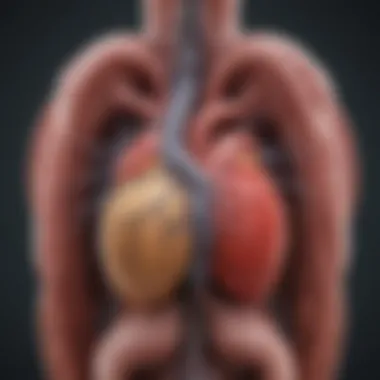

Common viral pathogens include Epstein-Barr Virus, Cytomegalovirus, and Human Immunodeficiency Virus. The significance of these pathogens arises from their prevalence in diverse populations and their potential role in triggering inflammatory responses that lead to pericardial effusion. These viruses are known for their capacity to invade cardiac tissue or the pericardial area, promoting fluid accumulation due to immune system response. The ability of these viruses to remain latent also presents a unique challenge in both diagnosis and management when symptoms manifest only after extended periods.
Mechanisms of Infection
The mechanisms by which these viruses cause pericardial effusion can involve direct invasion of the myocardial or pericardial tissues. Another component is the systemic inflammatory response triggered by viral infections, which may lead to increased vascular permeability. This permeability can facilitate fluid movement into the pericardial cavity. Understanding these mechanisms enhances the ability of healthcare professionals to recognize signs early and implement strategies to mitigate the complications associated with viral infections.
Bacterial Infections
Types of Bacterial Agents
Bacterial infections contributing to pericardial effusion often include organisms such as Streptococcus pneumoniae, Staphylococcus aureus, and Mycobacterium tuberculosis. Each bacterial agent possesses distinct pathogenic features that can lead to pericardial involvement. For instance, Streptococcus pneumoniae tends to cause more acute presentations, while Mycobacterium tuberculosis often results in a chronic effusion. Recognizing these agents helps in tailoring antibiotic therapies effectively, especially in cases where rapid intervention is critical.
Clinical Implications
The clinical implications of bacterial infections are considerable. These infections can lead to significant complications, including cardiac tamponade, which is life-threatening. Prompt recognition and treatment of bacterial pericarditis are therefore crucial in preventing adverse outcomes. Exploring these implications allows clinicians to develop comprehensive management plans that encompass both immediate treatment and potential long-term monitoring.
Fungal and Parasitic Infections
Less Common Pathogens
Fungal and parasitic infections are less common causes of pericardial effusion. Pathogens such as Histoplasma capsulatum and Toxoplasma gondii may not be top of mind compared to viral or bacterial agents, but their role is nonetheless critical. These pathogens can induce an inflammatory response, resulting in fluid collection. Their less frequent occurrence might mislead healthcare providers, which can delay diagnosis and treatment.
Associated Risks
The associated risks of these infections include delayed diagnosis due to their rarity. This delay can exacerbate the condition, leading to more severe effusions or complications. Patients with compromised immune systems, such as those with HIV/AIDS, are at higher risk for these infections. Educating medical professionals regarding the potential for fungal and parasitic infections as causes of pericardial effusion retains importance for effective patient monitoring and management.
Non-Infectious Causes
Understanding non-infectious causes of pericardial effusion is critical for a comprehensive approach to diagnosis and treatment. These causes represent a significant portion of cases and are diverse in origin, making it essential to identify them accurately. Non-infectious factors can lead to significant morbidity and further complications, impacting the management strategies adopted by healthcare professionals. Addressing these causes highlights the need for careful evaluation of a patient's medical history, underlying conditions, and systemic factors that could influence fluid buildup in the pericardium.
Autoimmune Diseases
Autoimmune diseases are a primary contributor to non-infectious pericardial effusion. These conditions cause the body's immune system to attack its own tissues, leading to inflammation. The complexity of autoimmune mechanisms makes it essential to understand how they can trigger fluid accumulation in the pericardial cavity.
Systemic Lupus Erythematosus
Systemic Lupus Erythematosus (SLE) is a chronic autoimmune disease known for its diverse range of symptoms. One specific aspect relevant to pericardial effusion is the associated inflammation that SLE can induce in the pericardium. A key characteristic of SLE is its propensity to affect multiple organ systems simultaneously, making fluid buildup in the pericardial space more likely. This connection is particularly beneficial to this article as it emphasizes SLE's role in diverse presentations of pericardial effusion.
A unique feature of SLE is its variable course, where patients may experience periods of exacerbation and remission. This fluctuation can complicate the management of pericardial effusion, as treatment may need adjustment alongside changes in disease activity.
Rheumatoid Arthritis
Rheumatoid Arthritis (RA) also plays a role in non-infectious pericardial effusion. This autoimmune disorder primarily affects the joints, but it can lead to systemic inflammation that impacts the heart. A key characteristic of RA is the chronic inflammatory state it creates, which may result in effusion formation over time. This aspect underscores the importance of recognizing RA as a relevant contributor to pericardial effusion.
A unique feature of RA is that it often affects individuals at a younger age compared to other chronic conditions. This early onset can complicate long-term outcomes, as the presence of pericardial effusion in younger patients may signal more serious underlying issues or increase morbidity related to cardiovascular health.
Malignancies
Malignancies encompass various forms of cancer, and they are significant causes of pericardial effusion, particularly in advanced stages. Understanding malignancy types and their specific pathways contributes to a clearer picture of fluid accumulation.
Primary Tumors
Primary tumors directly affecting the heart or surrounding structures can lead to pericardial effusion. These tumors are often associated with local invasion or inflammation, which can disrupt normal fluid dynamics. The key characteristic of primary tumors is their ability to alter the pericardial environment through physical mass effects or secretions that promote effusion development.
Highlighting primary tumors in this article is beneficial, as it demonstrates how localized cancer processes can lead to significant cardiac complications. A unique feature of primary tumors is their potential for rapid progression, which may necessitate acute intervention to manage the effusion and mitigate related symptoms.
Metastatic Disease
Metastatic disease is when cancer spreads from its original location to other parts of the body, including the pericardial space. This aspect is critical, as it often indicates advanced disease and can signify a poor prognosis. A key characteristic of metastatic disease is the variety of cancer types that may invade the pericardium. This diversity makes it an important focus of this article, highlighting the need for awareness of multiple disease states.
The unique feature of metastatic disease in this context is its systemic nature. Patients with metastatic cancer may present with effusion as a generalized complication rather than an isolated issue, emphasizing the importance of comprehensive evaluation in these cases.
Trauma and Injury
Trauma and injury are often overlooked causes of pericardial effusion. Understanding the types of trauma that can lead to fluid accumulation is essential for clinicians assessing patients with suspected effusions.
Blunt and Penetrating Trauma
Both blunt and penetrating trauma can result in pericardial effusion through different mechanisms. Blunt trauma can lead to direct injury to the heart or blood vessels, causing blood to accumulate in the pericardium. A key characteristic of this type of trauma is the immediate impact it has, as it can result in acute symptoms that require urgent evaluation.


Penetrating trauma introduces a risk of infection and can lead to significant effusions if not managed promptly. Highlighting this aspect in the article is beneficial to grasp the urgent nature of these cases and the need for rapid diagnostic imaging and interventions.
Post-Surgical Effusions
Post-surgical effusions can arise following cardiac or thoracic surgeries. They may result from the body's healing response or complications linked to the surgical procedure. A key characteristic of post-surgical effusions is their relatively high incidence in cardiovascular patients, highlighting the need for monitoring after procedures.
This section's unique feature is that post-surgical effusions can sometimes resolve spontaneously, yet may also require management, thus complicating the overall care strategy for patients recovering from surgery.
Chronic Conditions
Chronic conditions play a significant role in the development of pericardial effusions. Understanding these conditions provides vital insights into long-term management strategies.
Renal Failure
Renal failure can lead to pericardial effusion due to fluid overload and the inability of the kidneys to excrete excess fluids. A key characteristic is the systemic volume overload that can compromise normal pericardial dynamics. This connection is crucial for the article as it emphasizes the interplay between kidney function and cardiovascular health.
The unique feature of renal failure is its gradual progression, which can mean that effusions develop over time rather than acutely. This aspect highlights the importance of regular monitoring in patients with renal issues.
Heart Failure
Heart failure is another chronic condition that can significantly contribute to pericardial effusion. The fluid retention commonly associated with heart failure impacts the pericardial sac. A major characteristic of heart failure is the impaired pumping mechanism, which can lead to a backlog of pressure and fluid.
Addressing heart failure in this article shows its importance in understanding cardiac-related effusions. The unique feature of heart failure is its prevalence among various populations, making it a common consideration in patients with established heart conditions.
Idiopathic Pericardial Effusion
Idiopathic pericardial effusion is a pertinent subject within the broader discussion of fluid accumulation in the pericardial cavity. It bears significance due to the challenges it presents in diagnosis and treatment. Unlike cases with clear etiological factors, idiopathic effusion represents a category where the underlying cause remains elusive. This ambiguity complicates the management strategies for healthcare providers, as treatment often relies on understanding the root of the problem.
Definition and Frequency
Idiopathic pericardial effusion is defined as the unexplained accumulation of fluid in the pericardial cavity where no discernible reason can be determined. This condition can occur in isolation or alongside other cardiac issues. Epidemiological data suggests that idiopathic cases are observed in a significant proportion of individuals diagnosed with pericardial effusion. Estimates vary, but it is commonly reported that approximately 30% to 50% of all pericardial effusions are idiopathic in nature.
Researchers believe that this high percentage may be due in part to a limited understanding of certain pathophysiological mechanisms that lead to fluid accumulation. Furthermore, the absence of conducive laboratory or imaging findings makes diagnosis particularly difficult. Thus, idiopathic cases mandate a careful approach focusing on exclusion of other possible causes before a definitive classification can be made.
Research on Idiopathic Cases
Current research into idiopathic pericardial effusion often seeks to uncover potential underlying conditions that may have previously gone unnoticed. Studies have attempted to correlate idiopathic effusion with various autoimmune conditions, genetic predispositions, and environmental factors. For instance, there is an ongoing evaluation of factors that may contribute, such as viral infections that were unrecognized during earlier assessments.
Moreover, there is a push for greater utilization of advanced imaging techniques to capture subtler signs of pathology that could inform the diagnosis. Enhanced echocardiography, MRI scans, and innovative blood tests may provide the insights necessary for more accurate classification of these effusions. The aim of this research is to shift the perception of idiopathic cases from a diagnostic dead-end to a pathway towards understanding the complexities of the pericardial disease spectrum.
"Idiopathic pericardial effusion presents unique challenges and highlights the need for further investigation to unlock the mysteries of unexplained fluid accumulation."
The pursuit of knowledge in this field is essential for the development of improved treatment options, as well as for establishing a clearer connection between idiopathic effusion and other health conditions. Continuous exploration and a multidisciplinary perspective could eventually lead to enhanced patient outcomes.
Diagnostic Approaches
The diagnostic approaches to pericardial effusion serve as a crucial component of understanding this condition. These strategies encompass a range of techniques aimed at identifying the presence, extent, and potential underlying causes of fluid accumulation in the pericardial cavity. Reliable diagnostics are essential for appropriate management and treatment of the patient, as they inform clinical decision-making.
Medical Imaging Techniques
Echocardiography
Echocardiography is a primary imaging tool in evaluating pericardial effusion. This technique uses sound waves to create images of the heart, allowing healthcare professionals to visualize the pericardium and any fluid present. A key characteristic of echocardiography is its non-invasive nature, making it a beneficial option in most clinical settings.
A unique aspect of echocardiography is its ability to provide real-time assessment. This allows for immediate observations of hemodynamic changes that may occur due to the effusion. Moreover, echocardiography can distinguish between simple effusions and those that may pose more significant risks, such as tamponade.
However, it does have limitations. The quality of images can depend on the patient's body habitus and operator experience. In some cases, detecting small effusions or complex pericardial diseases may be challenging. Nevertheless, echocardiography remains a crucial first-line diagnostic tool due to its rapid availability and considerable advantages in assessing cardiac function.
CT Scans and MRI
CT scans and MRI have emerged as valuable adjuncts in the diagnostic process for pericardial effusion. A significant advantage of these imaging modalities is their ability to provide detailed cross-sectional views of the thoracic structures. This is particularly beneficial for better characterization of effusions and understanding the surrounding anatomy.
CT scans can quickly identify the presence of large pericardial effusions and associated complications. Their rapid acquisition time makes them practical in emergency settings. On the other hand, MRI offers superior soft tissue contrast, allowing for a detailed evaluation of both the pericardium and any underlying conditions that might contribute to the effusion. One unique feature of MRI is its capacity to assess myocardial inflammation, providing insights into potential causes.
Despite their strengths, these imaging techniques also have limitations. CT scans expose patients to ionizing radiation, which is a consideration in management, especially for younger individuals. MRI, while providing excellent detail, can be less accessible and time-consuming.
Laboratory Tests
Laboratory tests contribute indispensable information in the diagnostic process of pericardial effusion. They aid in understanding the underlying cause and the nature of the fluid accumulation.
Inflammatory Markers
Inflammatory markers are vital to identifying underlying processes that might lead to pericardial effusion. Common tests include C-reactive protein (CRP) and erythrocyte sedimentation rate (ESR) measurements, which signal the presence of inflammation within the body. A notable characteristic of inflammatory markers is their ability to indicate conditions, such as autoimmune diseases, that could be driving the effusion.
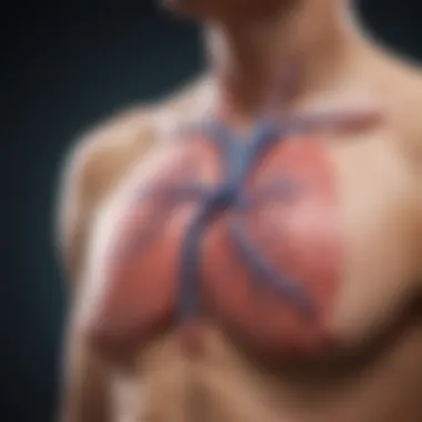

These tests can be easily obtained and are relatively inexpensive, making them practical for use in diverse clinical settings. However, one must consider that while elevated inflammatory markers imply an inflammatory process, they do not pinpoint the specific etiology of the effusion itself.
Culture Techniques
Culture techniques are instrumental in diagnosing infectious causes of pericardial effusion. They involve obtaining fluid samples through procedures like pericardiocentesis and analyzing them for the presence of pathogens. One of the key characteristics of culture techniques is their specificity; they can provide definitive evidence of bacterial, fungal, or viral infections when positive.
This method is especially beneficial in cases where infectious etiologies are suspected, as it guides targeted therapy. The main drawback, however, is the time required for cultures to grow and identify pathogens, which may delay treatment.
Treatment Options
Understanding the treatment options for pericardial effusion is vital for patients and healthcare providers. Treatment selection depends on the underlying cause and severity of the effusion. The aim is to relieve symptoms, prevent complications, and address any underlying conditions. Proper management of pericardial effusion can lead to significantly improved patient outcomes and quality of life.
Medical Management
Medications
Medications play a key role in managing pericardial effusion. Anti-inflammatory drugs such as non-steroidal anti-inflammatory drugs (NSAIDs) and corticosteroids are commonly used. Their primary function is to reduce inflammation in the pericardium, which can decrease fluid accumulation. The characteristic of these medications lies in their ability to provide symptomatic relief and possibly revert some conditions causing the effusion. They are typically a popular choice because they are often effective with manageable side effects.
The unique feature of these medications is their versatility. They can address multiple potential causes, from infectious processes to autoimmune conditions. However, the disadvantages include potential gastrointestinal side effects and the risk of immunosuppression with prolonged corticosteroid use, which warrants careful monitoring.
Monitoring Approaches
Monitoring approaches are essential in the management of pericardial effusion. Regular follow-ups and imaging studies help track the size of the effusion and assess symptoms. This approach enables clinicians to make timely adjustments to treatment plans based on the patient’s status. It is a beneficial choice in ensuring comprehensive care, especially when the underlying cause is chronic or progressive.
The key characteristic of monitoring approaches is their proactive nature. Continuous assessment allows for identification of any complications early, including cardiac tamponade. The unique feature of such monitoring is its capacity to provide reassurance to patients and aid clinicians in making informed decisions. However, it can be resource-intensive and might require frequent patient visits, which can be a barrier for some.
Interventional Procedures
Pericardiocentesis
Pericardiocentesis serves as both a diagnostic and therapeutic tool in managing pericardial effusion. This procedure involves inserting a needle to drain fluid from the pericardial cavity. Its importance lies in its ability to provide immediate relief from symptoms such as chest pain and dyspnea caused by fluid pressure on the heart.
The characteristic of pericardiocentesis is its minimally invasive nature. It is often preferred as a first-line intervention because it can be performed quickly and usually does not require a long recovery time. The unique feature of this procedure is that it allows for analysis of the fluid, which can help identify the underlying cause of the effusion. However, disadvantages include the risk of complications such as bleeding, infection, or injury to the heart or lungs.
Pericardial Window Surgery
Pericardial window surgery is a more invasive option for treating persistent effusions, especially when fluid re-accumulates after pericardiocentesis. This procedure involves creating a small opening in the pericardium to allow continuous drainage of fluid.
The key characteristic of pericardial window surgery is its effectiveness in managing recurrent or complicated effusions. It provides a longer-term solution compared to pericardiocentesis. The unique feature of this surgical intervention is the ability to directly address the causative factors of fluid buildup, offering both symptomatic relief and a potential cure. However, the disadvantages include longer recovery times and inherent surgical risks, which must be considered before proceeding.
Proper treatment and monitoring of pericardial effusion can lead to significant improvements in patient health and well-being. Understanding these options helps in tailoring care to individual needs.
Prognostic Factors
Understanding prognostic factors in pericardial effusion is crucial for predicting patient outcomes and informing treatment decisions. These factors encompass various elements, including the underlying conditions that contribute to effusion and the patient's overall health profile. Assessing these aspects can help healthcare providers develop effective management plans, improving both short-term and long-term results for their patients.
Impact of Underlying Conditions
The etiology of pericardial effusion often includes a range of underlying medical conditions. The presence of diseases such as systemic lupus erythematosus or malignancies can significantly influence the prognosis. Patients with autoimmune disorders face unique challenges, as these conditions may cause recurrent effusions stemming from chronic inflammation. Similarly, those diagnosed with cancers may encounter complications from direct tumor invasion of the pericardium or treatment-related effects.
Monitoring these conditions allows clinicians to tailor interventions effectively. For example, a patient with advanced malignancy may experience a different fluid dynamics than one suffering from benign inflammatory conditions. Understanding the impact of these factors on prognosis aids in anticipating the need for procedures like pericardiocentesis or the implantation of a pericardial window, particularly in patients with higher risks of complications.
Long-term Follow-Up
Long-term follow-up for patients with pericardial effusion is essential for several reasons. First, the persistence of effusions or recurrent episodes can indicate worsening underlying pathology or ineffective treatment strategies. Regular check-ups involving imaging and clinical assessment enable healthcare practitioners to track the resolution. The duration and frequency of follow-up visits may vary based on the underlying cause and response to treatment.
Additionally, follow-up focuses on managing potential complications that may arise after treatment. Some patients might develop adhesions or constrictive pericarditis after drainage procedures. Continuous assessment allows for timely intervention to mitigate these risks.
This process is important for ensuring the patient's quality of life, facilitating early detection of changes in health status, and adapting treatment plans accordingly.
Long-term management is vital, as untreated or poorly monitored pericardial effusion can lead to significant cardiac complications.
Culmination
The conclusion of this article serves as a critical step in encapsulating the various elements surrounding pericardial effusion. It pulls together the complexities discussed throughout the text, helping to solidify the understanding of this medical condition. The importance of recognizing the myriad causes is central to effective diagnosis and treatment. Emphasizing both infectious and non-infectious aspects brings a comprehensive insight into how different factors can influence fluid accumulation in the pericardial cavity.
Summary of Causes and Implications
In examining the causes of pericardial effusion, it becomes clear that the interplay of infectious agents, autoimmune diseases, malignancies, and other contributing factors necessitates a thorough diagnostic approach. Each cause can lead to distinct clinical implications, influencing the symptoms and management of the condition. For instance, effusion related to infections might require antibiotic therapy, while autoimmune origins may need immunosuppressive treatments. Understanding these causes allows healthcare professionals to tailor their interventions effectively, improving patient outcomes.
Future Directions in Research
Future research in pericardial effusion is vital to elucidating the underlying mechanisms of fluid accumulation. Areas that warrant further exploration include the role of novel biomarkers in early identification, advanced imaging techniques to assess effusion dynamics, and the long-term outcomes of different treatment methods. Over time, as our understanding of the condition deepens, targeted therapies could emerge, transforming how pericardial effusions are managed clinically.
A systematic approach to studying pericardial effusion can enhance patient care and ultimately improve healthcare standards.
The need for ongoing research and improved diagnostic tools cannot be overstated, as they will provide significant benefits and considerations for future management of this condition.


